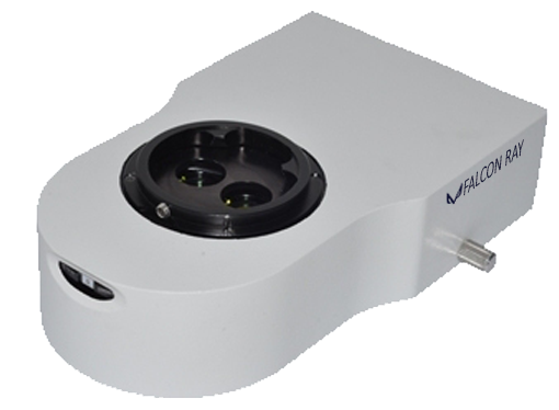
Upgrade any surgical microscope from Zeiss and Topcon to Leica and Haag-Streit, easily with simple removal of the binocular head and place the digitizer module in between. Facile calibration tool on system helps the installation user to adjust an in field and in focus image to what surgeon observes during the operation. Regardless of the live view, simultaneous framesets and images could be captured and sent to DICOM servers, as with their capability to be post processed and annotated. Real time image processing algorithms make the acquired visual data identical to what the surgeon perceives.

Surgical Microscopy Digital Upgrades
Falcon Ray provides digital upgrades to surgical microscopy to improve surgical practices with their educational, professional or marketing purposes.
Comply with Most Brands
Captures Exact Observation
Additional Vision to Human’s
Real Time Image Adjustment
Real Time Image Processing
High Clinical Ergonomy
Post Processing Tools
DICOM Export to PACS
Easy and Fast Installation
Standalone Functionality
Covering Surgical Applications
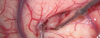
Neurology, Spine and Cranial
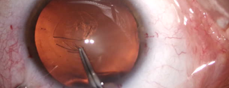
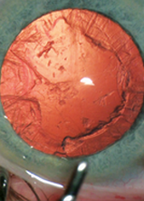
Ophthalmology
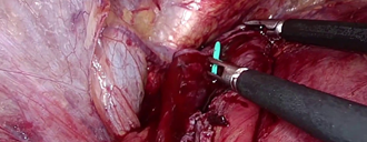
Urology and Nephrology
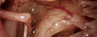
Otolaryngology (ENT)
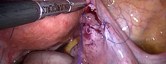
Gynecology
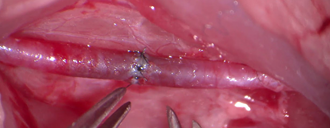
Plastic Recunstructive
Syncronized Digitization with Human Visual Understanding
When adopting digital imaging to archive and distribute the surgical observations from patients operations, not always what has been observed may be viewed in its digitized form. Falcon employs real time image processing and adjustment to push the digitized data as near as possible to clinicians observation in real time without surgical teams capability of employing an expert photographer. This encounters the reality in depth and tissue perception, as well as coloring and dynamic range.
Microscopes play a crucial role in day to day precision surgical practices. Stereo vision is a human factor that gives us the understanding of depth perception as with other hunter species known to ecology. Once however digital imaging is employed, a 2D image is reconstructed that would not synthesize a 3D vision as the human brain does. It is therefore important to make such an impression throughout the reconstructed image by the correct equation of aperture and focal length adjustment to reach the correct focus depth for surgical applications with respect to the magnification of operation. New solutions use polarized 3D Vision that may result visual data elimination.
Human visual understanding of luminescence and brightness encounters the value of pupil diameter along with retinal rod receptors and neuro-pulsation efficiency of brain. Otherwise, the clinician may be dealing with some extent of cataract that makes it hard to understand their luminous and brightness understanding. In digital imaging though, sensors quantum efficiency and optical elements including the adjustment lens and aperture play crucial roles in image brightness. However as long as Falcon Ray digital systems considers clinicians only tool to adjust the brightness to be their light source intensity tool, real time imaging algorithms are applied to image capture, in order to synchronize the digitized amplitude of data and dynamic range to cover all possible observations as an average human vision.

Any digital camera has an entity called the Dynamic Range, which is referred to the distance between the whitest white and the blackest black. This gray level is likewise applied to colors. When a camera sensor provides its image data to the Image Signal Processor of the camera, it is crucial for image processing algorithms to drive the sensor in a way that splits the dynamic range across the area in, where the required clinical data is present. Therefore it is crucial for microscope upgrades to capture the exact surgeon’s observation of colors by adjusting the right point for the true white in real time.
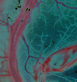
Once capturing the image or a frameset, the camera sensor could be driven in a way to enhance vision for what human eye is naked to. This could be imaging within the NIR frequency band as well as special image manipulations to reveal or project specific defects for surgeons to be extracted from the camera raw data. This gives the operation user a wider choice to illustrate any surgical action than the time a captured image or framset is processed. Sensor raw data is very much wider to navigate through than images with headers that could be only available for post processing options.
