Our Digital Radiography system provides the optimum clinical data in a compact solution with integrated detection and emission of X-Ray utilized by ultrafast realtime image processing. The post processing tools and DICOM export systems including DICOM Send to PACS, DICOM Print and burning a Standard DICOM Patient CD, will enable our devices data to be distributed in a standard approach when certain deffects are perfectly illustrated. With low dose imaging technology we protect our beloved ones as well as their Vets who are alternatively receiving ionizing radiation.

Clinical Radiography
With more than 14 years of experience in imaging via X-Ray, Falcon Ray provides state of the art digital X-Ray solutions for our beloved ones. Our vet imaging systems eliminate the patient to receive lower dose, once higher clinical data is acquired and a higher imaging ergonomy comes along with a higher imaging productivity.
Veterinary Digital Radiography (DR)
Emission and Detection Sync
High Frequency X-Ray Source
High Quantum Efficiency
Raltime Image Adjustment
Raltime Image Processing
DICOM Send to PACS
DICOM and Paper Print
Patient CD Generator
Post Processing Tools
High Clinical Ergonomy
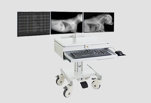
Veterinary Digital Fluoroscopy (DRF)
Suitable for surgical and scopic applications Falcon Ray Digital Fluoroscopy enables advanced paraclinical and surgical procedures to be exposed to vet’s eyes in real time and continously. Real time image processing algorithms make the tracing object and region of interest much observable in terms of the value of cliical data. This has been acheived by the integrated functionality of X-Ray emision and detection units through a single imaging co-ordination system. Excessive clinical data along with extremely low dose and high ergoomy in device operation makes it suitable for a diversity of vet applications.
Emission and Detection Sync
High Frequency X-Ray Source
High Quantum Efficiency
Raltime Image Adjustment
Raltime Image Processing
DICOM Send to PACS
DICOM and Paper Print
Patient CD Generator
Post Processing Tools
High Clinical Ergonomy
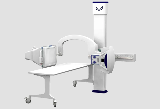
Integrated Emmission and Detection of X-Ray
Fro an X-Ray image to be captured or a Fluoroscopic study to be done with the least possible dose and highest clinical data, many factors are into consideration! From the power-electronics and photonics of an X-Ray source to quantum efficiency and micro-electronics of the X-Ray detector along with the real time image signal processing, all are required to meet the latest technology and standards.
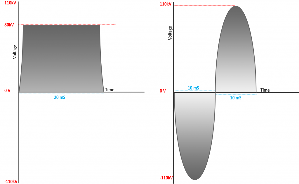
X-Ray sources being driven by high frequency power supplies or SMPS, are highly efficient in producing X-Ray as their emission remains constant during the exposure period. Low frequency x-ray sources in contrast, turn on and off for 100 to 120 times per second as the alternating current changes, where images experience the fogs caused by different levels of X-Ray energy and therefore noise on clinical data.
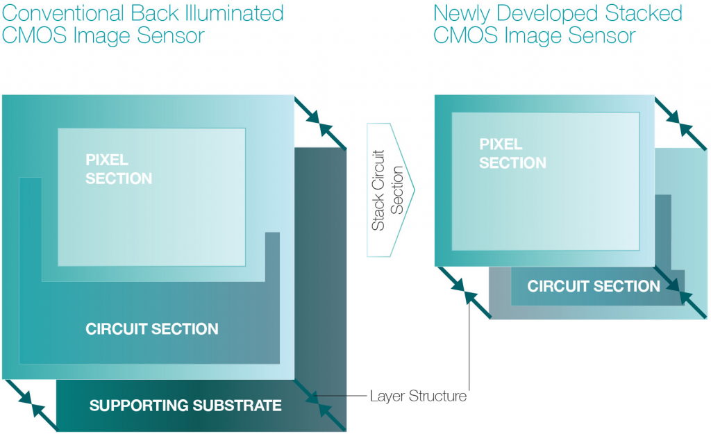
When increasing quantum efficiency of a detector, more signal to noise ratio, as well as a higher range of clinical data could be acquired. Falcon Ray to that end, has obtained back-illuminated CMOS sensor technology to optimize each photodiodes apperture to its widest. This would allow higher photon intensity to charge the diode. Otherwise the quantum efficiency is being enhanced with our CsI scintilators as well.

Any digital X-Ray detector has an entity called the Dynamic Range, which is referred to the distance between the whitest white and the blackest black. Gray level is likewise applied within this area. When a detector provides its image data to the Image Signal Processor of the radiography, it is crucial for image processing algorithms to drive the source and detector in a way that splits the dynamic range across the area, where the required clinical data exists. Therefore it is crucial for digital radiographies to capture the clinical data of interest by adjusting the right point for the true white in real time.
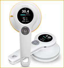
High frequency X-Ray sources being integrated to high quantum efficiency X-Ray detectors in Falcon Ray radiographic devices are the reason for which the equivalent dose could be extremely reduced. Meanwhile the Image Signal Processing make real time adjustments to the image, the priority would be with shortening the exposure time and intensity. Also the image processing algorithms driving the ISP navigate through the digital image, in order to split the dynamic range, where the clinical data is located. Our integrated colimation control plays a crucial role in dose reduction as well.
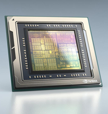
Regardless of how advance a radiography’s detector and X-Ray emitter be, the brain that make them work synchronized distinguish one from the other. Image Signal Processing has become advanced to intelligent signal processing of radiographic image acquisition once being responsible for the integration of the X-Ray source and detector. Dose reduction along with increasing the value of clinical data is the critical ability of Falcon Ray radiography systems that are utilized by advanced image processing and adjustment algorithms to manage acquiring a proper set of clinical data within the image.
